Fraunhofer researchers develop innovative method for clinical applications in the field of stem-cell-based retinal implants
Controlled manufacture, storage and freezing of artificial retinal cells
Age-related macular degeneration (AMD) is a degenerative eye condition that affects approximately ¼ of the population. Particularly in the western world, it is the most frequent cause of blindness in humans. In most cases, the chronic progression of the retinal condition is not curable, as the window of opportunity in which any therapeutic action can be taken is very small. One potential new form of treatment is presented by stem-cell therapy. Fraunhofer researchers have now developed a new method for the production and clinical application of stem-cell-based retinal implants, which could contribute towards the successful treatment of AMD. Their approach thereby is to replace damaged tissue with stem-cell-based transplants.
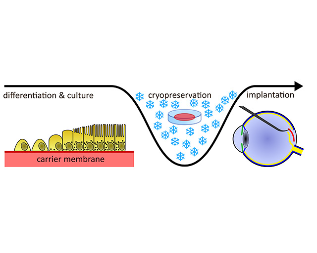
In addition to the further development of the manufacturing process, one of the greatest current challenges in working with human induced pluripotent stem cells (hiPSC) is the long-term storage and the transport of the cells. Within the framework of the “KryoRet” project, the Fraunhofer institutes for Biomedical Engineering IBMT, for Silicate Research ISC, and for Surface Engineering and Thin Films IST, together with the Fraunhofer Translational Center Regenerative Therapies TLC-RT, therefore investigated the technical and biotechnological prerequisites involved in enabling hiPSC-based retinal implants to be manufactured more efficiently and stored for longer periods. In this context, the design of the cryogenic container and the type of cryopreservation itself were of particular significance. A further important aspect within the project was the quality control of the transplant. The Fraunhofer scientists were provided with support by specialists from the Augenklinik Sulzbach/Saar (eye clinic in Sulzbach/Saar, Germany).
A custom-fit carrier membrane, which is manufactured in a laboratory, serves as the physiological and functional basic structure of the transplants. It consists of ORMOCER®, that is, inorganic-organic hybrid molecules, which are combined with silica gel based fibers in order to achieve the desired diffusion properties and, simultaneously, to ensure good adhesion of the retinal pigment epithelial (RPE) cells to the membrane. Only with sufficient adhesion can the functionality of the cells be guaranteed. The question as to how the adhesion of the cells to the membrane surface can be optimally controlled through plasma treatment was investigated at the Fraunhofer IST.
In total, it takes around 60 days for the complete assembly of the then implantation-ready tissue. Secure storage technologies for the artificial RPE cells are therefore necessary, in which the quality and vitality of the cells are preserved. For this purpose, the cells should be frozen in a cryogenic container in a controlled and gentle manner without any damage occurring to their structure. In order to achieve this, the researchers at the Fraunhofer IST experimented with a variety of layer formers. By means of a plasma jet, adhesive layers were applied locally in the cryogenic container, upon which particles accumulate which, in turn, serve as a nucleation agent for the phase transition from water to ice. One goal of the experiments was the specific control of the crystallization process of ice formation during freezing in the plastic container by means of coatings and the development of an optimal cryoprotocol.
At the same time, the quality of the cells must always be ensured. During the entire process, no damage can be permitted to the implant itself. At the Fraunhofer IST, investigations were therefore performed in order to determine the extent to which machine-learning methods can be utilized in a non-invasive image-based process for the evaluation of the RPE cells with regard to their quality and functionality. The image material necessary for training the AI with different development phases of the RPE cells in varying qualities was provided by the project partners, the Fraunhofer IBMT and the Fraunhofer ISC. Such methods of image evaluation could potentially be transferred to other application areas. At the core is software for AI-supported image evaluation, programmed within the framework of the development of a digital infrastructure at the Fraunhofer IST.
Last modified:
